| Catalog Number | Product | Size | Price | |
|---|---|---|---|---|
| A1039-200 | Anti-human DDR1 Antibody PRTH-101 | 200µg | $325 | Order |
| A1039-100 | Anti-human DDR1 Antibody PRTH-101 | 100µg | $185 | Order |
| Catalog Number | A1039 |
|---|---|
| Product Name | Anti-human DDR1 antibody PRTH-101 |
| Source | Recombinant anti-human DDR1 mAb produced from HEK293 cells |
| Clone | PRTH-101 (VH and VL sequences are derived from Liu J et al, 2023) |
| Species Reactivity | Human |
| Isotype | Human IgG1 |
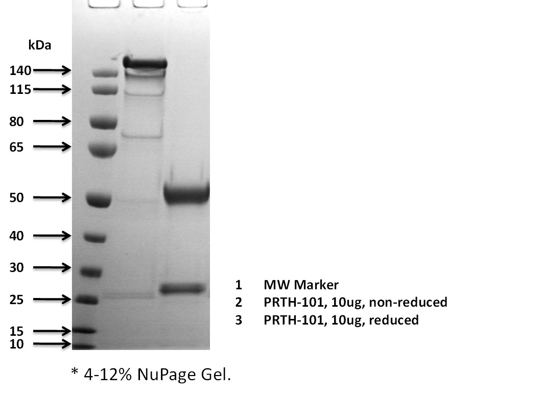
| Purity | >95% |
| Formulation | 50 mM Na Acetate, pH 5.2; Sterile |
| Stability & Storage | 1 month at 4oC; 12 months at <-20oC; Avoid repeated freeze-thaw |
| Protein Aggregation | Not obvious on SDS-PAGE |
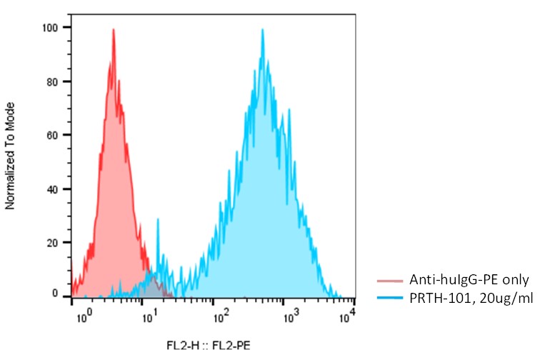
| Application | Flow cytometry, ELISA, cell-based assay |
| Product Datasheet: | Download PDF |
Detection of human DDR1 on DDR1-CHO-K1 cells (Cat. #C3030) by flow cytometry. Anti-human DDR1 antibody PRTH-101 (Cat. #A1039) was incubated with human DDR1-CHO-K1 cells (Cat. #C3030), followed by staining with PE-anti-human IgG.
DDR1 (discoidin domain receptor tyrosine kinase 1), also known as CD167, CAK, DDR, NEP, HGK2, PTK3, RTK6, TRKE, EDDR1, MCK10, NTRK4, and PTK3A, is a receptor tyrosine kinase (RTK) and belongs to a subfamily of tyrosine kinase receptors with a homology region to the Dictyosteliumdiscoideum protein discoidin I in its extracellular domain. DDR1 consists of three regions (an extracellular ligand binding domain, a transmembrane domain, and an intracellular region containing a kinase domain), with its kinase activity induced by receptor-specific ligand binding. Collagen binding to DDR1 stimulates its autophosphorylation, activating kinase activity and signaling to downstream signaling pathways. DDR1 expression is restricted to epithelial cells, particularly in the kidney, lung, gastrointestinal tract, and brain and is significantly over-expressed in several human tumors from breast, ovarian, esophageal, and brain. DDR1 plays a key role in the development and progression of breast and ovarian cancer and is a promising therapeutic target.
Johnson, J. D., Edman, J. C., Rutter, W. J., Proc. Nat. Acad. Sci. 90: 5677-5681, 1993.
Chen, L., et al, Frontiers in Cell and Dev. Bio. 9: 747314, 2021.
Letinger, B.,Int Rev Cell Mol Biol., 310: 39-87, 2014.
Vogel, W., et al., Mol. Cell, 1: 13–23, 1997.
Liu, J, et al, Journal for ImmunoTherapy of Cancer 2023;11:e006720. doi:10.1136/ jitc-2023-006720